Printable template
Parts of a Cell (Blank)
Cell diagram to label.
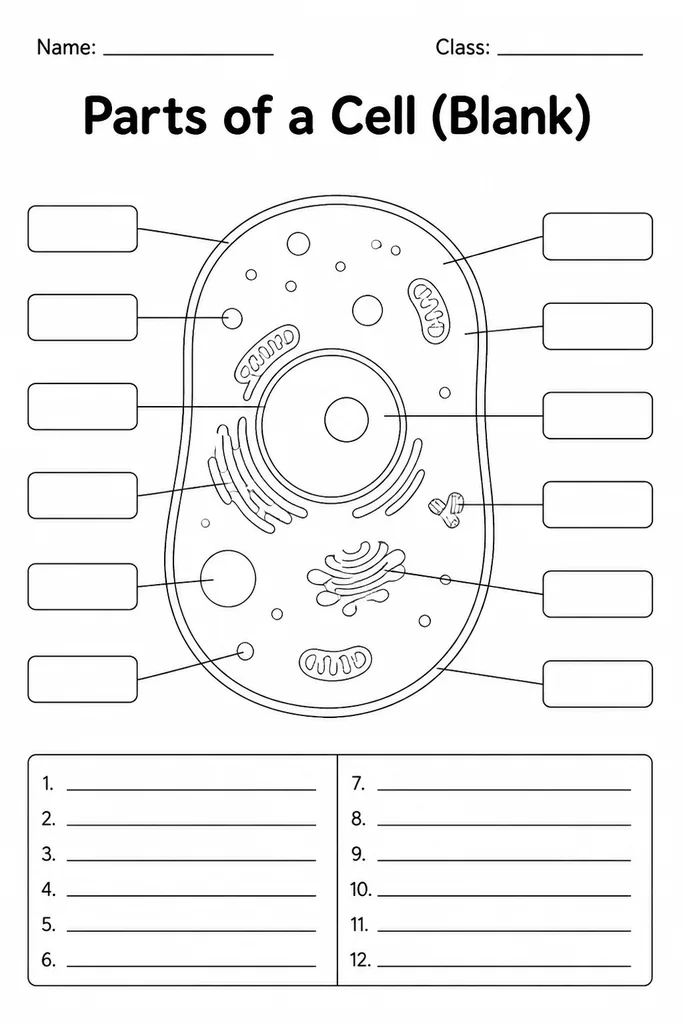
This blank cell diagram template provides a clean, unlabelled cross-section illustration suitable for both animal and plant cell studies in grades 5–8. Empty label lines radiate from each organelle, giving students space to write in structures such as the nucleus, mitochondria, cell membrane, endoplasmic reticulum, vacuole, and chloroplasts (plant cell version). Teachers use it at the start of a cell biology unit to activate prior knowledge, mid-unit for guided note-taking, and at the end as a no-prep quiz. Because the diagram ships without any text, it serves as a neutral assessment tool—students can't glean answers from partial labelling. The template is sized to fit a standard lab notebook page, making it practical for science interactive notebooks and binder inserts alike.
Learning objectives
- Identify and correctly name organelles in animal and plant cells
- Distinguish structural differences between plant and animal cell diagrams
- Practice using precise biological terminology when labelling
- Support note-taking during lectures or textbook reading
- Prepare for cell biology quizzes and standardised science tests
- Build familiarity with scientific diagram conventions
How to use this template
- Download the free PDF—choose the animal cell, plant cell, or both versions—and print on letter or A4 paper.
- Distribute during a cell biology lesson or as independent study material.
- Students label each organelle line using class notes, a textbook, or from memory for assessment.
- Review answers together using a projected answer-key diagram.
- Glue into science interactive notebooks or laminate for repeated dry-erase use.
Classroom & home ideas
- Project a microscope image of a real onion cell and ask students to map what they see onto the blank template.
- Run a 'label race'—partners take turns adding one organelle label at a time to a shared laminated sheet.
- Use the plant and animal versions side by side for a compare-and-contrast activity on cell differences.
- Assign as a flipped-learning task: students label at home before the teacher explains in class.
- Include in a science station rotation where students self-check with a sealed answer envelope.
Skills practised
Frequently asked questions
Does the template show a plant cell, an animal cell, or both?
KiwiBee offers both versions in the same download. The plant cell includes chloroplasts, a central vacuole, and a rigid cell wall; the animal cell shows centrioles and lacks a cell wall.
Which organelles are labelled lines provided for?
Lines point to the nucleus, nucleolus, cell membrane, cytoplasm, mitochondria, Golgi apparatus, endoplasmic reticulum (rough and smooth), ribosomes, lysosomes, vacuole, and—on the plant version—chloroplasts and cell wall.
Is this appropriate for grade 5 students who are seeing cells for the first time?
Yes. Pair it with a word bank printed below the diagram so first-time learners can focus on spatial placement rather than pure recall.
Can I shrink the diagram to fit a composition notebook?
Yes. In your print settings, choose 75–80% scale and it fits neatly on a standard composition-notebook page for interactive notebooks.
Likes & comments
See what other teachers think and share a helpful note.
Create more worksheets in our Worksheet Studio
Generate fresh worksheets for any grade, topic, and language — free.
Open the Worksheet StudioRelated worksheets
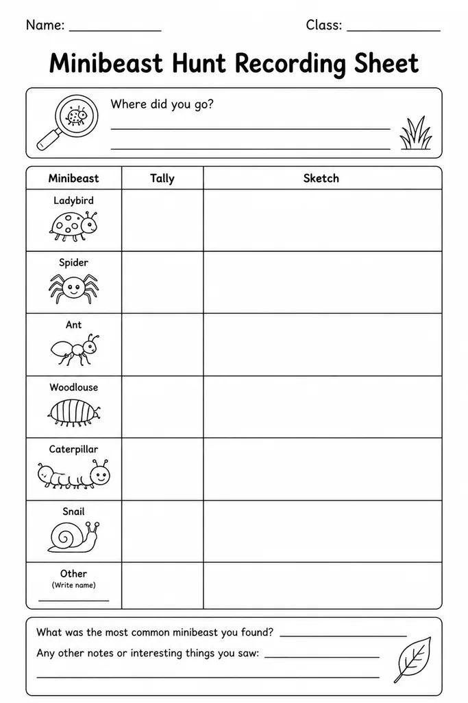
Minibeast Hunt Recording Sheet
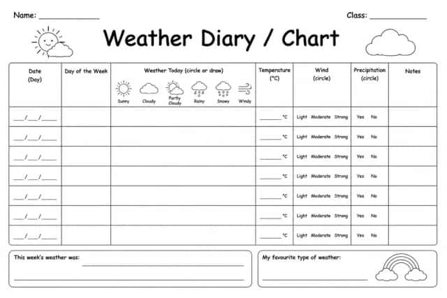
Weather Diary / Chart
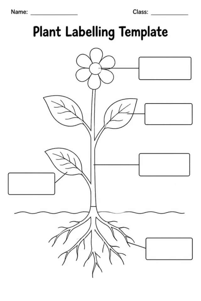
Plant Labelling Template
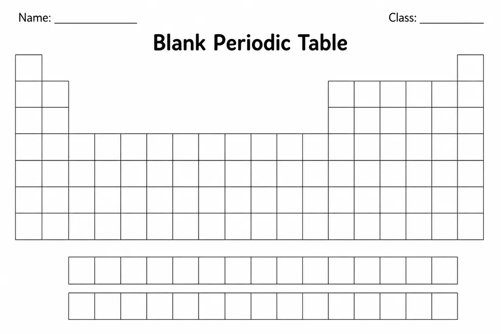
Blank Periodic Table
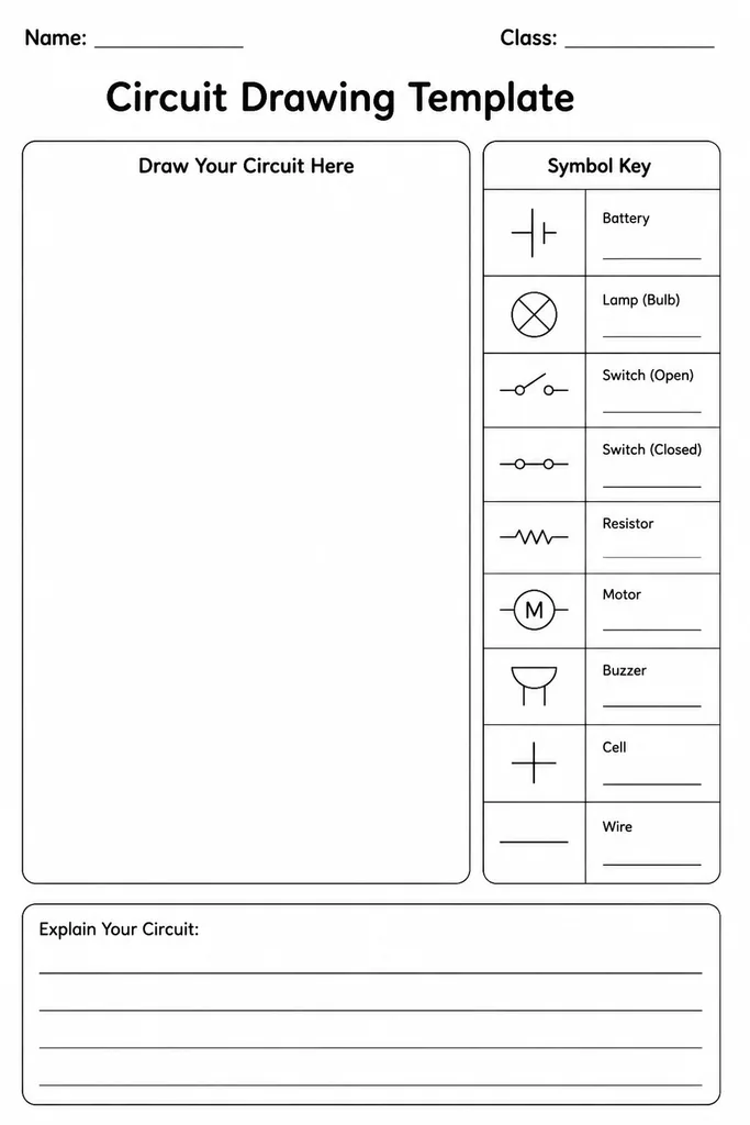
Circuit Drawing Template
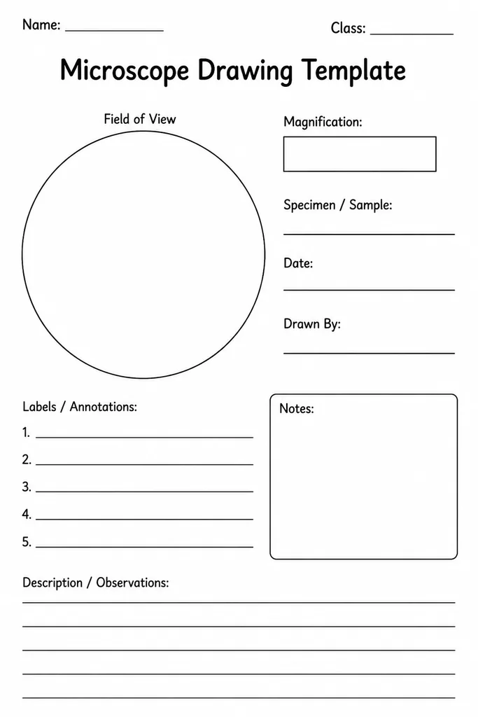
Microscope Drawing Template
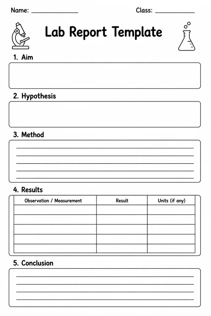
Lab Report Template