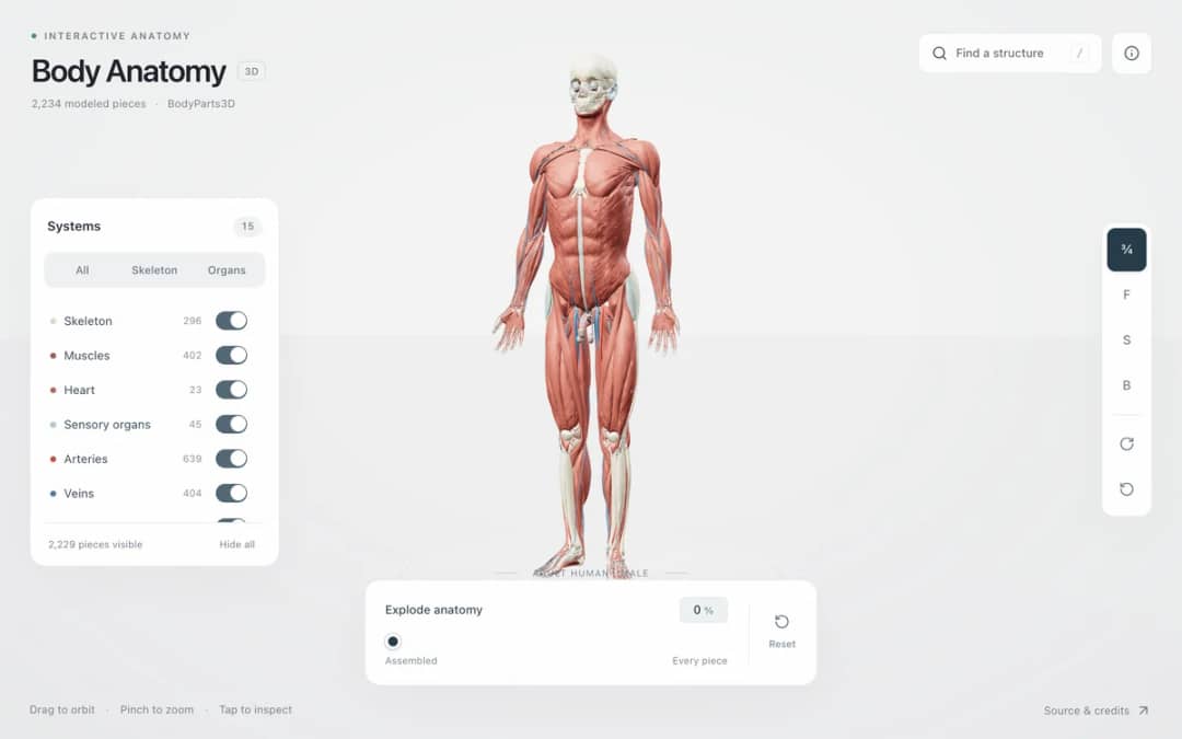
Explore an adult male reference body in 3D. Reveal 14 anatomical systems, search thousands of named structures, isolate organs, and pull 2,209 modeled pieces apart to see how the body is organized.
—
KiwiBee uses optional Google Analytics, Google Ads, and Meta Pixel cookies to understand adult visitors, measure completed sign-ups, and improve campaigns. Essential cookies always stay on. OpenAI’s ChatGPT Ads Pixel also measures page views and completed enquiries after advertising consent, and may use hashed contact details for matching. Privacy policy
This model shows a virus particle built from identical protein subunits tiled over an icosahedron, which has 20 faces, 12 vertices and 30 edges. Students pick a T number of 1, 4, 9 or 16 to retile the shell and watch the capsomer and subunit counts change. Sliders set the diameter and rotation, and toggles show spike proteins or a cutaway with the RNA genome inside. It is useful for talking about symmetry, why viruses reuse one small protein, and how structure relates to a tiny genome.
Yes. It is free and runs in any modern browser, including classroom whiteboards and tablets. No sign-up or installation is required.
A capsid is the protein shell that surrounds a virus's genome. Many viruses use an icosahedral shell made from 60 times T identical subunits, where T is the Caspar-Klug number.
Turn on the Cutaway + RNA core toggle. Part of the shell is removed so you can see the coiled RNA genome. You can also drag to rotate the model and scroll to zoom.
No. The spikes here are schematic, and real spike counts and positions differ between viruses. The model also shows only T = 1, 4, 9 and 16, not twisted lattices like T = 3 or 7.
Drop it into your LMS, blog, or class site — free.
<iframe src="https://kiwibee.io/en/simulations/virus-capsid-3d-simulation/embed" title="Virus Capsid | KiwiBee" loading="lazy" allowfullscreen style="width:100%;height:min(70vh,620px);min-height:420px;border:0;border-radius:16px"></iframe>Continue exploring
Explore an adult male reference body in 3D. Reveal 14 anatomical systems, search thousands of named structures, isolate organs, and pull 2,209 modeled pieces apart to see how the body is organized.
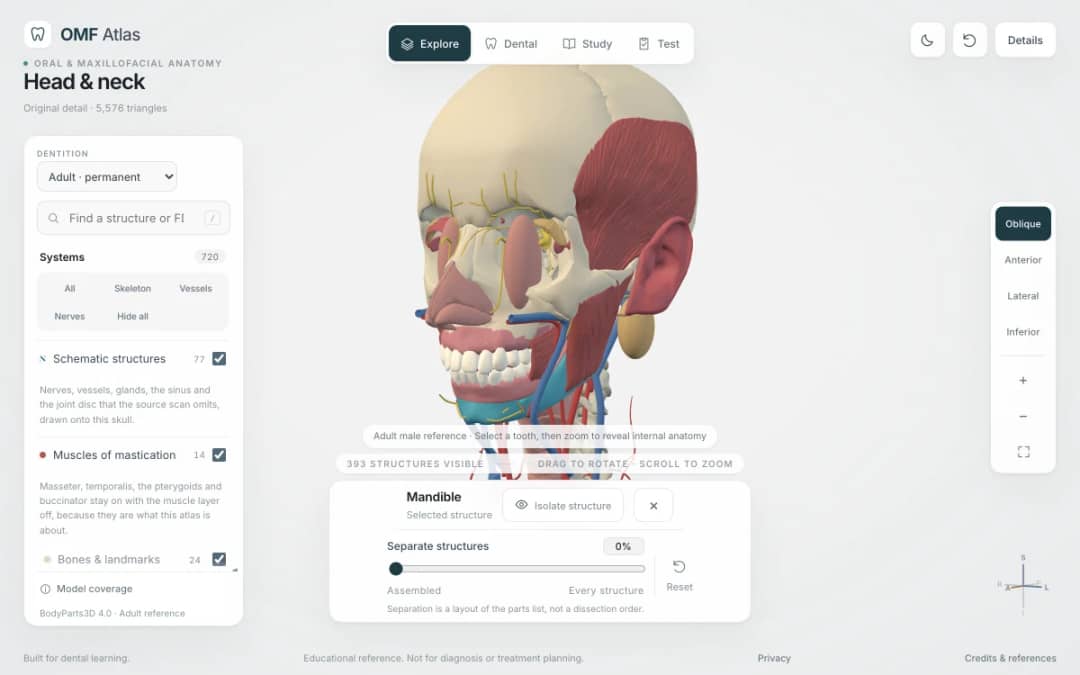
Explore the head and neck in 3D: 643 source meshes across 14 layers, 47 facial and masticatory muscles, and 77 drawn nerves, vessels and glands the source scan omits. Open any tooth onto its own pulp canal, and follow the seven stages of tooth development.

Free interactive photosynthesis simulation. Explore photosynthesis hands-on online — great for whole-class or independent practice.

Free interactive states of matter simulation. Explore states of matter hands-on online — great for whole-class or independent practice.

Free interactive shadows simulation. Explore shadows hands-on online — great for whole-class or independent practice.
From model to evidence
Use a worksheet to capture predictions, observations, and explanations after students explore.
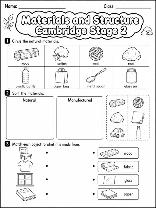
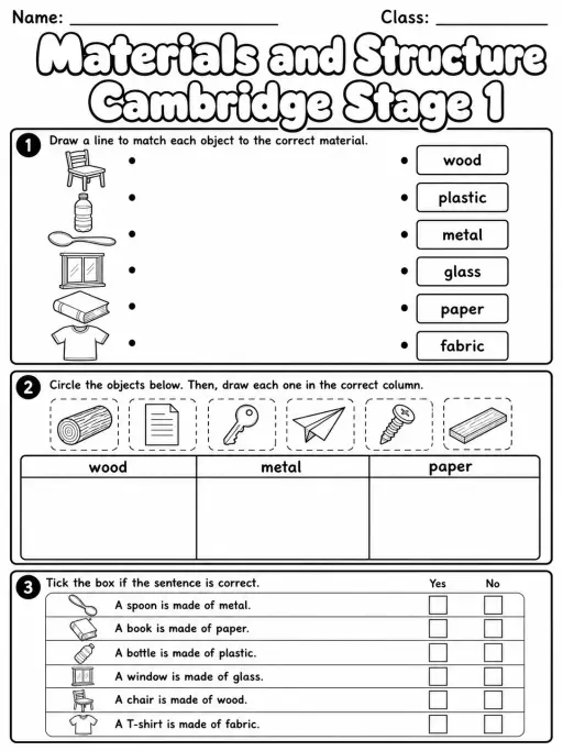
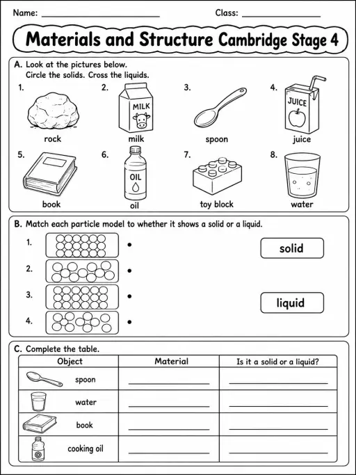
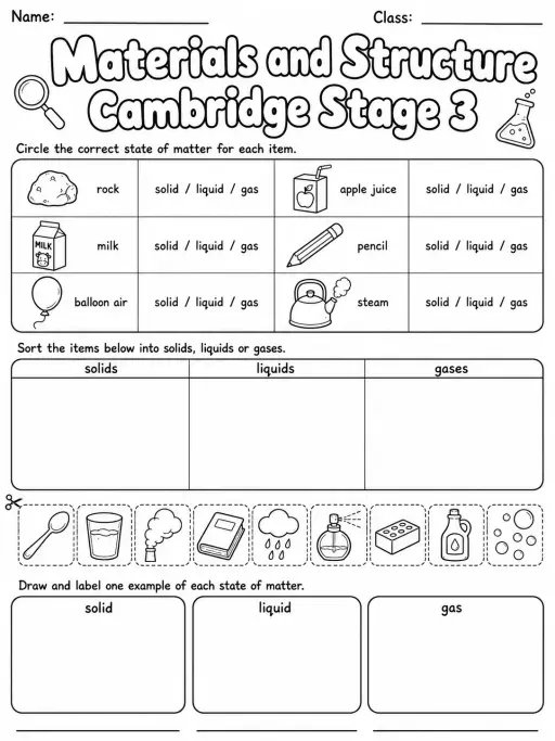
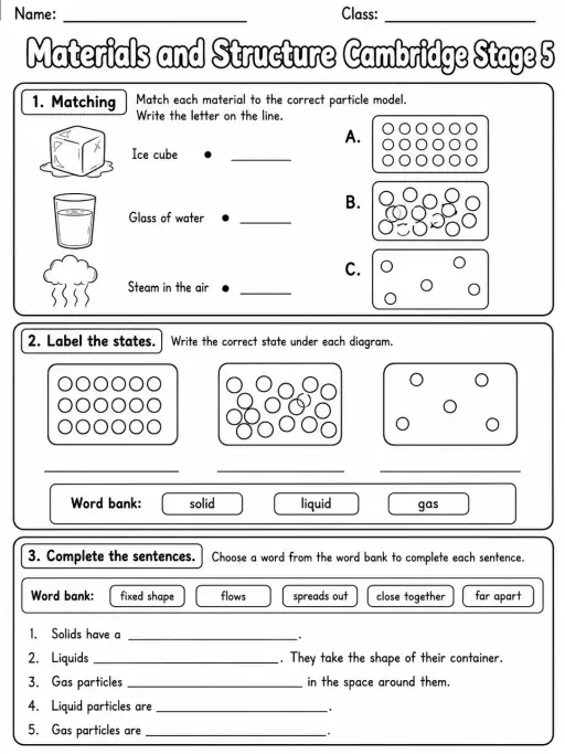
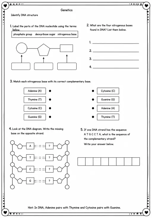
KiwiBee's free teacher workspace brings lesson planning, worksheets, classroom tools, and gradebook together in one place.
Start free and create your first class Home
 All solutions
 Pricing
 About us
 KiwiBee for Non-profits
 Events & webinars
 For big organisations
 For schools
 For centers
 For team leaders
 For teachers
 AI and resources
 Resources
 Printable worksheets
 Lesson Packs
 Interactive simulations
 Teacher tools
 AI studio
 Ideas and research