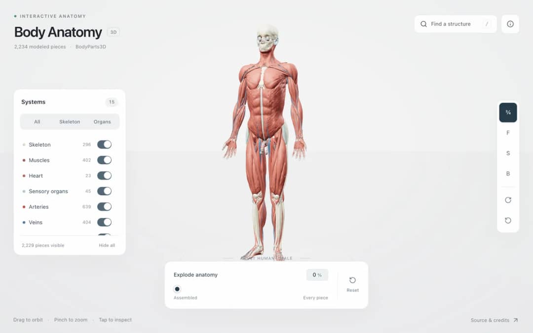
Explore an adult male reference body in 3D. Reveal 14 anatomical systems, search thousands of named structures, isolate organs, and pull 2,209 modeled pieces apart to see how the body is organized.
—
KiwiBee uses optional Google Analytics, Google Ads, and Meta Pixel cookies to understand adult visitors, measure completed sign-ups, and improve campaigns. Essential cookies always stay on. OpenAI’s ChatGPT Ads Pixel also measures page views and completed enquiries after advertising consent, and may use hashed contact details for matching. Privacy policy
Muscle Sarcomere Contraction 3D demonstrates the sliding filament mechanism of skeletal muscle physiology. Framed between two blue Z-disc plates, thin green actin filaments overlap a central thick red myosin filament studded with hinged myosin heads. Students can drag either Z-disc horizontally or click the myosin heads to trigger a calcium release pulse. Sliders for Calcium (Ca2+), ATP concentration, and external Load force allow students to observe active cross-bridge cycling, measure sarcomere shortening from 2.5 micrometers down to 1.6 micrometers, and see why depleting ATP locks cross-bridges in the rigor state.
No. Neither the thin actin filaments nor the thick myosin filaments change length. Instead, myosin heads ratchet along the actin filaments, pulling the two Z-discs closer together and increasing filament overlap.
At rest, tropomyosin strands cover the binding sites on actin. When an action potential triggers Ca2+ release from the sarcoplasmic reticulum, Ca2+ binds troponin and rolls tropomyosin out of the way so myosin heads can grab actin.
A fresh ATP molecule must bind to the myosin head for it to detach from actin after a power stroke. Without ATP, the myosin cross-bridges remain permanently locked to actin, making the muscle rigid.
Drop it into your LMS, blog, or class site — free.
<iframe src="https://kiwibee.io/en/simulations/sliding-filament-muscle-3d-3d-simulation/embed" title="Muscle Sarcomere Contraction | KiwiBee" loading="lazy" allowfullscreen style="width:100%;height:min(70vh,620px);min-height:420px;border:0;border-radius:16px"></iframe>Continue exploring
Explore an adult male reference body in 3D. Reveal 14 anatomical systems, search thousands of named structures, isolate organs, and pull 2,209 modeled pieces apart to see how the body is organized.
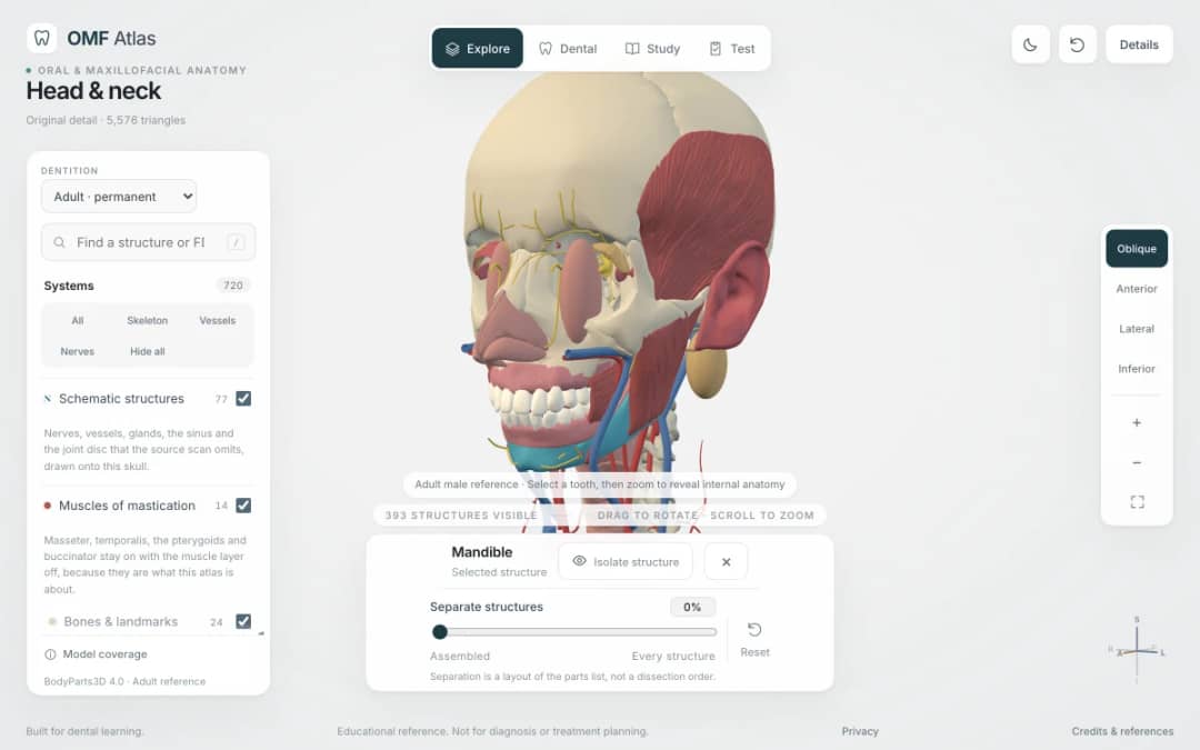
Explore the head and neck in 3D: 643 source meshes across 14 layers, 47 facial and masticatory muscles, and 77 drawn nerves, vessels and glands the source scan omits. Open any tooth onto its own pulp canal, and follow the seven stages of tooth development.

Free interactive photosynthesis simulation. Explore photosynthesis hands-on online — great for whole-class or independent practice.

Free interactive states of matter simulation. Explore states of matter hands-on online — great for whole-class or independent practice.

Free interactive shadows simulation. Explore shadows hands-on online — great for whole-class or independent practice.
KiwiBee's free teacher workspace brings lesson planning, worksheets, classroom tools, and gradebook together in one place.
Start free and create your first class Home
 All solutions
 Pricing
 About us
 KiwiBee for Non-profits
 Events & webinars
 For big organisations
 For schools
 For centers
 For team leaders
 For teachers
 AI and resources
 Resources
 Printable worksheets
 Lesson Packs
 Interactive simulations
 Teacher tools
 AI studio
 Ideas and research