Oral & Maxillofacial Atlas
Science · Oral and Maxillofacial Anatomy · High School
关于这个模拟实验
Oral & Maxillofacial Atlas is a head-and-neck 3D anatomy explorer built on BodyParts3D geometry. Students can rotate and zoom the assembly, reveal or hide 14 anatomical layers, search and isolate 643 named source structures, separate the whole model into a parts inventory, open a tooth onto a sectioned 3D cutaway, and work through the stages of tooth development. It is a dental-education reference, not a diagnostic or treatment-planning tool.
学生可以探索什么
- Reveal and hide 14 layers of the head and neck, including 47 facial and masticatory muscles registered from the BodyParts3D 3.0 archive.
- Show 77 schematic structures the source dataset does not contain — the trigeminal and facial nerves, the external carotid tree, the parotid gland and duct, the maxillary sinus and the temporomandibular disc — or switch the whole set off.
- Search a structure by name or FDI number, isolate it, and separate the assembly into an ordered parts inventory.
- Zoom into a tooth to open its 3D cutaway, move the section plane, and toggle enamel, dentin, pulp and root canals, cementum, ligament, alveolar bone and gingiva.
- Follow the seven stages of odontogenesis, the eruption chronology for both dentitions, and the anomalies that arise at each stage.
学习目标
- 1Name the bones, muscles, nerves, vessels and glands of the head and neck and locate them relative to one another in three dimensions.
- 2Relate the external form of a tooth to its internal anatomy, its root canal arrangement and its supporting periodontium.
- 3Sequence the stages of tooth development and connect each stage to the tissues it forms and the anomalies that arise from it.
- 4Distinguish a structure segmented from a scan from one drawn schematically, and say what each kind of model can and cannot show.
教师常问的问题
Is the Oral & Maxillofacial Atlas free to use?
Yes. It runs in a modern browser with no sign-up or download required, and it loads no third-party resources.
What anatomy does the model represent?
The head and neck of the BodyParts3D 4.0 adult male reference, cropped above the lower neck: 643 source meshes across 14 layers. Seventy-seven further structures — most of the nerves, vessels, glands and the joint disc an oral and maxillofacial atlas needs — are absent from that dataset and are drawn instead, fitted to landmarks measured on the licensed meshes. They are labelled as schematic wherever they are named.
Can students look inside a tooth?
Yes. Selecting a tooth and zooming in opens a rotatable 3D cutaway on a movable section plane, with enamel, dentin, pulp and root canals, cementum, ligament, alveolar bone and gingiva each switchable. The teeth come from a published cone beam CT dataset, so a canal is shown as the scan resolved it, not as an apical working length.
Who is it for?
It was built for dental students and dental professionals, and the anatomical depth reflects that. In school it suits senior biology and health-science students studying the skull, the jaw and the teeth; it is not pitched at primary or lower-secondary classes.
嵌入这个模拟实验
免费嵌入你的学习平台、博客或班级网站。
<iframe src="https://kiwibee.io/zh-cn/simulations/omf-atlas-simulation/embed" title="Oral & Maxillofacial Atlas | KiwiBee" loading="lazy" allowfullscreen style="width:100%;height:min(70vh,620px);min-height:420px;border:0;border-radius:16px"></iframe>继续探索
更多互动科学模型
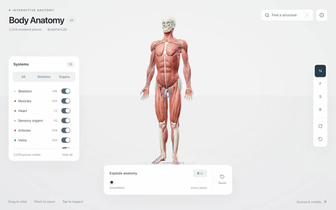
Explore an adult male reference body in 3D. Reveal 14 anatomical systems, search thousands of named structures, isolate organs, and pull 2,209 modeled pieces apart to see how the body is organized.

Free interactive photosynthesis simulation. Explore photosynthesis hands-on online — great for whole-class or independent practice.

Free interactive weather simulation. Explore weather hands-on online — great for whole-class or independent practice.

Free interactive shadows simulation. Explore shadows hands-on online — great for whole-class or independent practice.

Free interactive states of matter simulation. Explore states of matter hands-on online — great for whole-class or independent practice.
把互动科学带进你的课堂
KiwiBee 的免费教师工作台把备课、工作表、课堂工具和成绩册集中在一处。
免费开始,创建你的第一个班级 For big organisations
 For schools
 For centers
 For team leaders
 For teachers
 AI and resources
 可打印的工作表
 交互式模拟
交互式模拟 教师工具
教师工具 AI 工作室
 思想和研究
思想和研究